About
Selected publications
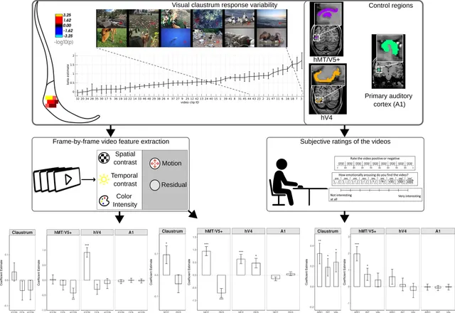
- The Human Visual Claustrum Responses to Physical Stimulus Properties and Subjective Content During Movie Viewing
Abstract
The human claustrum is a small bilateral grey matter structure that is highly interconnected with cortical and subcortical regions. It has been implicated in different functions including sleep, multisensory integration, consciousness and attention, yet its exact function remains unclear. The primate claustrum is known to have distinct sensory regions, with the visual zone recently demonstrated in humans using high-resolution fMRI. In this study, we investigated stimulus properties that drive human visual claustrum activity. First, we tested the association of its response with various low- and mid-level physical stimulus features, including temporal and spatial contrast, color and motion. Second, we tested the association with subjective ratings of arousal, valence and interest. To compare the claustrum's responses with visual cortical regions, we performed the same analysis with the hV4 and hMT/V5+ complex. We found that the claustrum's visual response was associated with motion, as well as with arousal, interest and valence. The pattern of claustrum responses was similar to hMT/V5+. Given the well-established link between arousal and attentional allocation, as well as between saliency and motion, our results suggest that the visual claustrum may contribute to saliency detection and attention modulation during the sensory input.
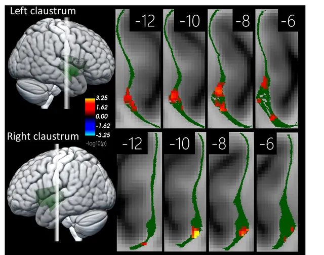
InHuman Brain Mapping - High-resolution 7T fMRI reveals the visual zone of the human claustrum
Abstract
The claustrum is a thin grey matter structure located between the insular cortex and the putamen. The function of the claustrum is largely unknown with diverse hypotheses ranging from multisensory integration and consciousness to attention and cognitive control. Much research on the function of the claustrum relies on invasive techniques in animal models, as the claustrum’s uniquely thin shape makes it difficult to image non-invasively in human subjects. In the current proof-of-concept study, we used high-resolution ultra-high field (7 Tesla) functional magnetic resonance imaging (fMRI) to measure activity in the human claustrum during the processing of naturalistic stimuli. We presented short video clips as visual only, auditory only, or audiovisual conditions while participants performed a central fixation task. We found distinct visual responses in both the left and the right claustrum at a consistent spatial location across participants, hemispheres, and sessions. We also found deactivations in response to auditory stimulation. These deactivations were confined to the right claustrum and did not overlap with visual activity. The deactivation in response to auditory stimulation demonstrates the complexity of the claustrum’s functional organization and suggests functional differentiation within the claustrum. This is the first study to demonstrate sensory-specific effects within the human claustrum. It opens the possibility for studying the claustrum’s role in higher-level aspects of sensory processing in humans.
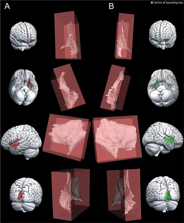
InImaging Neuroscience - High-resolution dataset of manual claustrum segmentation
Abstract
The claustrum has a unique thin sheet-like structure that makes it hard to identify in typical anatomical MRI scans. Attempts have been made to identify the claustrum in anatomical images with either automatic segmentation techniques or using atlas-based approaches. However, the resulting labels fail to include the ventral claustrum portion, which consists of fragmented grey matter referred to as “puddles”. The current dataset is a high-resolution label of the whole claustrum manually defined using an ultra-high resolution postmortem MRI image of one individual. Manual labelling was performed by four independent research trainees. Two trainees labelled the left claustrum and another two trainees labelled the right claustrum. For every hemisphere we created a union of the two labels and assessed the label correspondence using dice coefficients. We provide size measurements of the labels in MNI space by calculating the oriented bounding box size. These data are the first manual claustrum segmentation labels that include both the dorsal and ventral claustrum regions at such a high resolution in standard space. The label can be used to approximate the claustrum location in typical in vivo MRI scans of healthy individuals.
InData in Brief